Physicochemical characterization and in vitro biocompatibility assessment of hydrothermally synthesized hydroxyapatite from oyster shells
DOI:
https://doi.org/10.55713/jmmm.v36i4.2657Keywords:
Hydroxyapatite, Oyster shells, Hydrothermal, Biocompatibility, Saos-2Abstract
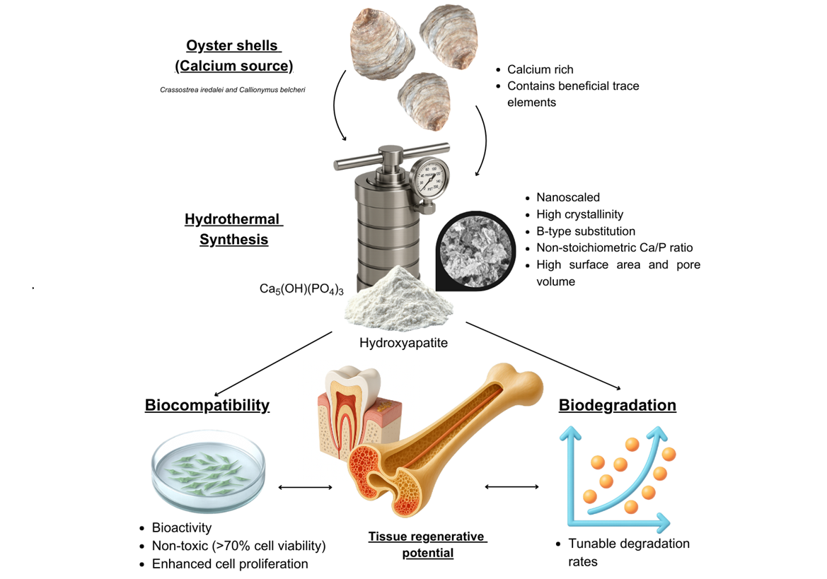
Naturally produced Hydroxyapatite (HAP) yields beneficial attributes with regards to its biological functions including cell compatibility, proliferation and osteoinduction. This study synthesized HAP from oyster shells as a calcium source with multiple hydrothermal reaction times including 1 h, 6 h and 24 h at 150℃. A wet precipitated control sample was produced at room temperature. Characterization using FTIR, XRD, SEM-EDX and BET were conducted, followed by a degradation analysis. Bio-compatibility study of Oys-HAP was performed against Saos-2 osteoblast-like cells. The results showed nano scaled elongated and oval-shaped Oys-HAP particles with high crystallinity, beta-type carbonated, non-stoichiometric Ca/P ratio with a maximum surface area of 41.39 m2·g‒1. Furthermore, Oys-HAP samples were non-toxic and significantly enhanced Saos-2 cell proliferation by 65.6% compared to the negative control. These findings demonstrate the potential of oyster shell-derived Oys-HAP as a sustainable and effective biomaterial for biomedical applications.
Downloads
References
H. Gul, M. Khan, and A. S. Khan, “Bioceramics,” in Handbook of Ionic Substituted Hydroxyapatites, Elsevier, 2020, pp. 53–83. DOI: https://doi.org/10.1016/B978-0-08-102834-6.00003-3
P. Murray, “Biocompatibility of biomaterials for dental tissue repair,” in Biocompatibility of Dental Biomaterials, Elsevier, 2017, pp. 41–62. DOI: https://doi.org/10.1016/B978-0-08-100884-3.00004-7
R. P. K. Penttinen, “Cell interaction with bioactive glasses and ceramics,” in Bioactive Glasses, Elsevier, 2018, pp. 145–180. DOI: https://doi.org/10.1016/B978-0-08-100936-9.00009-5
K. M. Galler, F. P. Brandi, S. Kirchhof, and M. Widbiller, “Suitability of different natural and synthetic biomaterials for dental pulp tissue engineering,” Tissue Engineering Part A, vol. 24, no. 3–4, pp. 234–244, 2017. DOI: https://doi.org/10.1089/ten.tea.2016.0555
M. Sadat-Shojai, M.-T. Khorasani, and A. Jamshidi, “Hydro-thermal processing of hydroxyapatite nanoparticles—A Taguchi experimental design approach,” Journal of Crystal Growth, vol. 361, pp. 73–84, 2012. DOI: https://doi.org/10.1016/j.jcrysgro.2012.09.010
G. Zhang, J. Chen, S. Yang, Q. Yu, Z. Wang, and Q. Zhang, “Preparation of amino-acid-regulated hydroxyapatite particles by hydrothermal method,” Materials Letters, vol. 65, no. 3, pp. 572–574, 2011. DOI: https://doi.org/10.1016/j.matlet.2010.10.078
S. C. Wu, H. C. Hsu, W. H. Wu, and W. F. Ho, “Enhancing bioactivity and mechanical properties of nano-hydroxyapatite derived from oyster shells through hydrothermal synthesis,” Nanomaterials (Basel), vol. 14, no. 15, 2024. DOI: https://doi.org/10.3390/nano14151281
R. A. Bapat, C. P. Joshi, P.Bapat, T. V. Chaubal, R. Pandurangappa, N. Jnanendrappa, B. Gorain, S. Khurana, and P. Kesharwani, “The use of nanoparticles as biomaterials in dentistry,” Drug Discovery Today, vol. 24, no. 1, pp. 85–98, 2019. DOI: https://doi.org/10.1016/j.drudis.2018.08.012
M. Motskin, D. M. Wright, K. Muller, N. Kyle, T. G. Gard, A. E. Porter, and J. N. Skepper, “Hydroxyapatite nano and micro-particles: Correlation of particle properties with cytotoxicity and biostability,” Biomaterials, vol. 30, no. 19, pp. 3307–3317, 2009. DOI: https://doi.org/10.1016/j.biomaterials.2009.02.044
S. C. Wu, H. C. Hsu, H. F. Wang, S. P. Liou, and W. F. Ho, “Synthesis and characterization of nano-hydroxyapatite obtained from eggshell via the hydrothermal process and the precipitation method,” Molecules, vol. 28, no. 13, 2023. DOI: https://doi.org/10.3390/molecules28134926
C. M. Mardziah, S. Ramesh, M. F. Abdul Wahid, H. Chandran, A. Sidhu, S. Krishnasamy, and J. Purbolaksono, “Effect of zinc ions on the structural characteristics of hydroxyapatite bio-ceramics,” Ceramics International, vol. 46, no. 9, pp. 13945–13952, 2020. DOI: https://doi.org/10.1016/j.ceramint.2020.02.192
E. Landi, A. Tampieri, G. Celotti, and S. Sprio, “Densification behaviour and mechanisms of synthetic hydroxyapatites,” Journal of the European Ceramic Society, vol. 20, no. 14–15, pp. 2377–2387, 2000. DOI: https://doi.org/10.1016/S0955-2219(00)00154-0
S. Ibrahim, S. A. Bakar, N. S. K. Bashah, N. M. Zainon, S. Sabudin, A. Seeni, O. J. Ping, and N. Yahaya, “Cytocompatible carbonate apatite derived from gypsum granules for bone regeneration,” Malaysian Journal of Medicine and Health Sciences, vol. 21, pp. 73–77, 2025.
T. Kokubo and H. Takadama, “How useful is SBF in predicting in vivo bone bioactivity?,” Biomaterials, vol. 27, no. 15, pp. 2907–2915, 2006. DOI: https://doi.org/10.1016/j.biomaterials.2006.01.017
Biological evaluation of medical devices Part 5: Tests for in vitro cytotoxicity , ISO 10993-5:2009, Aug. 09, 2025. [Online]. Available: https://www.iso.org/standard/36406.html.
Biological evaluation of medical devices Part 12: Sample preparation and reference materials, ISO 10993-12:2021, Aug. 09, 2025. [Online]. Available: https://www.iso.org/ standard/75769.html.
S. Ramesh, C. Y. Tan, R. Tolouei, M. Amiriyan, J. Purbolaksono, I. Sopyan, and W. D. Teng, “Sintering behavior of hydroxyapatite prepared from different routes,” Materials & Design, vol. 34, pp. 148–154, 2012. DOI: https://doi.org/10.1016/j.matdes.2011.08.011
S.-C. Wu, H.-C. Hsu, L.-C. Ou, and W.-F. Ho, “Effects of hydro-thermal temperature on the synthesis and characterization of bioactive hydroxyapatite nanoparticles from oyster shell,” Journal of the Australian Ceramic Society, vol. 61, no. 4, pp. 1525‒1536, 2025. DOI: https://doi.org/10.1007/s41779-025-01189-w
V. R. Sivaperumal, R. Mani, M. S. Nachiappan, and K. Arumugam, “Direct hydrothermal synthesis of hydroxyapatite/alumina nano-composite,” Materials Characterization, vol. 134, pp. 416–421, 2017. DOI: https://doi.org/10.1016/j.matchar.2017.11.016
A. Yelten-Yilmaz and S. Yilmaz, “Wet chemical precipitation synthesis of hydroxyapatite (HA) powders,” Ceramics International, vol. 44, no. 8, pp. 9703–9710, 2018. DOI: https://doi.org/10.1016/j.ceramint.2018.02.201
C. Chappard, G. André, M. Daudon, and D. Bazin, “Analysis of hydroxyapatite crystallites in subchondral bone by Fourier transform infrared spectroscopy and powder neutron diffraction methods,” Comptes Rendus Chimie, vol. 19, no. 11–12, pp. 1625–1630, 2016, DOI: https://doi.org/10.1016/j.crci.2015.03.015
T. J. Webster, C. Ergun, R. H. Doremus, R. W. Siegel, and R. Bizios, “Enhanced osteoclast-like cell functions on nanophase ceramics,” Biomaterials, vol. 22, no. 11, pp. 1327–1333, 2001. DOI: https://doi.org/10.1016/S0142-9612(00)00285-4
A. Pataquiva Mateus, F. J. Monteiro, and M. Pía Ferraz, “Nano-particles of hydroxyapatite: Preparation, characterization and cellular approach - An overview,” Revista Mutis, vol. 3, no. 2, pp. 43–56, 2013. DOI: https://doi.org/10.21789/22561498.884
J. P. Lafon, E. Champion, and D. Bernache-Assollant, “Processing of AB-type carbonated hydroxyapatite Ca10−x(PO4)6−x(CO3)x (OH)2−x−2y(CO3)y ceramics with controlled composition,” Journal of the European Ceramic Society, vol. 28, no. 1, pp. 139–147, 2008. DOI: https://doi.org/10.1016/j.jeurceramsoc.2007.06.009
A. I. Bucur, R. A. Bucur, Z. Szabadai, C. Mosoarca, and P. A. Linul, “Influence of small concentration addition of tartaric acid on the 220°C hydrothermal synthesis of hydroxyapatite,” Materials Characterization, vol. 132, pp. 76–82, 2017. DOI: https://doi.org/10.1016/j.matchar.2017.07.047
T. Tolga Demirtaş, G. Kaynak, and M. Gümüşderelioğlu, “Bone-like hydroxyapatite precipitated from 10×SBF-like solution by microwave irradiation,” Materials Science and Engineering: C, vol. 49, pp. 713–719, 2015. DOI: https://doi.org/10.1016/j.msec.2015.01.057
R. Z. LeGeros, “Calcium phosphate-based osteoinductive materials,” Chemical Reviews, vol. 108, no. 11, pp. 4742–4753, 2008, DOI: https://doi.org/10.1021/cr800427g
I. R. Gibson, and W. Bonfield, “Novel synthesis and characterization of an AB-type carbonate-substituted hydroxyapatite,” Journal of Biomedical Materials Research, vol. 59, no. 4, pp. 697–708, 2002. DOI: https://doi.org/10.1002/jbm.10044
K. Ariga, A. Vinu, Y. Yamauchi, Q. Ji, and J. P. Hill, “Nano-architectonics for mesoporous materials,” Bulletin of the Chemical Society of Japan, vol. 85, no. 1, pp. 1–32, 2012. DOI: https://doi.org/10.1246/bcsj.20110162
W. Liang, Y. Niu, S. Ge, S. Song, J. Su, and Z. Luo, “Effects of hydrothermal treatment on the properties of nanoapatite crystals,” International Journal of Nanomedicine, vol. 7, pp. 5151–5158, 2012. DOI: https://doi.org/10.2147/IJN.S34077
L. Yin, X. Xu, C. Chu, P. Lin, H. Huang, B. Luo, and C. Yang, “In-vitro characterization and evaluation of mesoporous titanium dioxide composite hydroxyapatite and its effectiveness in occluding dentine tubules,” BMC Oral Health, vol. 22, Art. no. 43, 2022. DOI: https://doi.org/10.1186/s12903-021-01989-z
P. Habibovic, M. C. Kruyt, M. V. Juhl, S. Clyens, R. Martinetti, L. Dolcini, N. Theilgaard, and C. A. van Blitterswijk, “Comparative in vivo study of six hydroxyapatite-based bone graft substitutes,” Journal of Orthopaedic Research, vol. 26, no. 10, pp. 1363–1370, 2008. DOI: https://doi.org/10.1002/jor.20648
M. Bohner, “Calcium orthophosphates in medicine: From ceramics to calcium phosphate cements,” Injury, vol. 31, pp. D37–D47, 2000. DOI: https://doi.org/10.1016/S0020-1383(00)80022-4
N. Patel, I. R. Gibson, S. Ke, S. M. Best, and W. Bonfield, “Calcining influence on the powder properties of hydroxyapatite,” Journal of Materials Science: Materials in Medicine, vol. 12, no. 2, pp. 181–188, 2001. DOI: https://doi.org/10.1023/A:1008986430940
M. Rouahi, E. Champion, O. Gallet, A. Jada, and K. Anselme, “Physico-chemical characteristics and protein adsorption potential of hydroxyapatite particles: Influence on in vitro biocompatibility of ceramics after sintering,” Colloids and Surfaces B: Biointerfaces, vol. 47, no. 1, pp. 10–19, 2006. DOI: https://doi.org/10.1016/j.colsurfb.2005.11.015
N. Suresh, V. K. Sweety, N. Suresh, A. R. Suraj, T. Waltimo, and S. Anil, “NATURAL sources of hydroxyapatite for bio-medical applications,” Ceramics International, vol. 52, no. 2, pp. 1383–1391, 2025. DOI: https://doi.org/10.1016/j.ceramint.2025.12.019
Y. Ebisawa, T. Kokubo, K. Ohura, and T. Yamamuro, “Bioactivity of CaO·SiO2-based glasses:in vitro evaluation,” Journal of Materials Science: Materials in Medicine, vol. 1, no. 4, pp. 239–244, 1990. DOI: https://doi.org/10.1007/BF00701083
Y. Wang, W. He, H. Hao, J. Wu, and N. Qin, “Eggshell derived Se-doped HA nanorods for enhanced antitumor effect and curcumin delivery,” Journal of Sol-Gel Science and Technology, vol. 87, no. 10, pp. 600–607, 2018. DOI: https://doi.org/10.1007/s10971-018-4765-0
J. Malešič, M. Kadivec, M. Kunaver, T. Skalar, and I. K. Cigić, “Nano calcium carbonate versus nano calcium hydroxide in alcohols as a deacidification medium for lignocellulosic paper,” Heritage Science, vol. 7, no. 1, p. 50, 2019. DOI: https://doi.org/10.1186/s40494-019-0294-6
W. E. Olds, D. C. W. Tsang, and P. A. Weber, “Lignite-derived humic substances for treatment of acid mine drainage,” Environmental Engineering Science, vol. 30, no. 10, pp. 638–645, 2013. DOI: https://doi.org/10.1089/ees.2012.0412
S. Ahn, S. Lee, and S. Lim, “Antimicrobial properties of thermally processed oyster shell powder for use as calcium supplement,” Foods, vol. 14, no. 15, p. 2579, 2025. DOI: https://doi.org/10.3390/foods14152579
R. Nithya, and N. Meenakshi Sundaram, “Biodegradation and cytotoxicity of ciprofloxacin-loaded hydroxyapatite-polycapro-lactone nanocomposite film for sustainable bone implants,” International Journal Nanomedicine, vol. 10, no. Sup 2, pp. 119–127, 2015. DOI: https://doi.org/10.2147/IJN.S79995
V. Ganesan, M. Devaraj, S. K. Govindan, V. S. Kattimani, and G. Easwaradas Kreedapathy, “Eggshell derived mesoporous biphasic calcium phosphate for biomedical applications using rapid thermal processing,” International Journal of Applied Ceramic Technology, vol. 16, no. 5, pp. 1932–1943, 2019. DOI: https://doi.org/10.1111/ijac.13270
S. Palakurthy, P. A. Azeem, K. Venugopal Reddy, V. Penugurti, and B. Manavathi, “A comparative study on in vitro behavior of calcium silicate ceramics synthesized from biowaste resources,” Journal of the American Ceramic Society, vol. 103, no. 2, pp. 933–943, 2019. DOI: https://doi.org/10.1111/jace.16745
S. Palakurthy, V. G. R. K, R. K. Samudrala, and A. A. P, “In vitro bioactivity and degradation behaviour of β-wollastonite derived from natural waste,” Materials Science and Engineering: C, vol. 98, pp. 109–117, 2019. DOI: https://doi.org/10.1016/j.msec.2018.12.101
E. M. Czekanska, M. J. Stoddart, J. R. Ralphs, R. G. Richards, and J. S. Hayes, “A phenotypic comparison of osteoblast cell lines versus human primary osteoblasts for biomaterials testing” Journal of Biomedical Materials Research Part A, vol. 102, no. 8, pp. 2636–2643, 2014. DOI: https://doi.org/10.1002/jbm.a.34937
Downloads
Published
How to Cite
Issue
Section
Categories
License
Copyright (c) 2026 Journal of Metals, Materials and Minerals

This work is licensed under a Creative Commons Attribution-NonCommercial-NoDerivatives 4.0 International License.
Authors who publish in this journal agree to the following terms:
- Authors retain copyright and grant the journal right of first publication with the work simultaneously licensed under a Creative Commons Attribution License that allows others to share the work with an acknowledgment of the work's authorship and initial publication in this journal.
- Authors are able to enter into separate, additional contractual arrangements for the non-exclusive distribution of the journal's published version of the work (e.g., post it to an institutional repository or publish it in a book), with an acknowledgment of its initial publication in this journal.